(BMJ) - A 49-yo woman presents w/ proximal myopathy and purple striae on torso and thighs. PMHx: DM, HTN, hypokalemia. Meds: insulin, metformin, 2 BP meds. Later she reveals using a skin-whitening moisturizer and topical clobetasol. What is the diagnosis?
Scleroderma
Elephantiasis
Lichen sclerosus
Cushing syndrome
Metabolic syndrome
You are correct. The patient had Cushing syndrome secondary to exogenous corticosteroid use. She had been applying a steroid-containing skin whitener + topical 0.05% clobetasol for the past 10 yrs. A morning cortisol was suppressed at 11 nmol/L (ref: 171-536 nmol/L). Two months later, off all corticosteroids, morning cortisol and ACTH were normal, and she was off insulin and antihypertensives.
(BMJ) – A post-op patient admitted to the ICU complained of acute pain and paresthesia in the first 3 digits of her left hand seconds after aspiration of blood for ABG from a radial artery catheter. What is the diagnosis?
Buerger disease
Raynaud phenomenon
Frostbite
Arterial air embolism
Vitiligo
You are correct. This patient acquired an arterial air embolism during radial artery catheterization during arterial blood gas analysis. The nurse immediately noticed that air had been entrained in the tubing and removed the line promptly. The patient’s signs and symptoms resolved spontaneously within a few minutes.
(BMJ) – A 32-yo male presented with a 2-mo Hx of fatigue, 4-kg weight loss, and dyspepsia. PMHx: none. Labs: mild normocytic anemia. Upper endoscopy: unusual raised lesion in 2nd part of duodenum. What is the diagnosis?
Duodenal ulcer
Protrusion of ampulla of Vater
Metastatic melanoma
Retained bile
Crohn disease
You are correct. Histology and immune studies confirmed metastatic melanoma. CT showed lung, adrenal, and cerebral mets, but skin exam was normal. Histological review of a nevus excised 6 yrs prior revealed a subtle melanoma, not appreciated originally. GI involvement occurs in 60% of metastatic melanoma pts w/ only 4% diagnosed before death. Initial presentation w/ duodenal mets is unusual; typical sx may include pain, bleeding, and obstruction.
(BMJ) - A 3-wk-old boy born via uncomplicated spontaneous vaginal delivery at 39 wks presented with a 1-wk Hx of facial pustules. Prenatal care did not start until 22 wks gestation. The infant was otherwise well-appearing and did not seem bothered by the rash. What is it?
Atopic dermatitis
Herpes simplex
Neonatal pustular melanosis
Group A streptococcus
Acne neonatorum
You are correct. Acne neonatorum, also known as neonatal cephalic pustulosis, is a benign, self-limited rash that presents shortly after birth. Acneiform papules and pustules are located on the cheeks, forehead, and, sometimes, chest. Herpes lesions, in contrast, are vesicular. Group A strep infection is usually associated with signs of sepsis. Neonatal pustular melanosis typically affects the entire body including palms and soles. Atopic dermatitis is often itchy and doesn't present until after 3 mos of life.
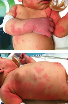
(BMJ) - An 18-day-old baby with normal birth hx presented with a new rash of sharply demarcated, red, irregularly-shaped papules and plaques. The baby appeared well w/o lymphadenopathy or organomegaly. Blood work confirmed the diagnosis. What is it?
Allergic reaction to diaper cream
Neonatal psoriasis
Tinea corporis
Herpesvirus 6-associated urticaria multiforme
Staphylococcal scalded skin syndrome
You are correct. Real-time PCR for viral DNA revealed human herpesvirus 6 (HHV-6) infection; other serological tests and cultures were negative. Three days after admission, the skin lesions completely resolved. Antibiotic therapy was stopped and patient was discharged. At 1-mo follow-up, HHV-6 DNA had decreased and the infant remained well without recurrence of rash.
(BMJ) - A 21-yo man on azathioprine for Crohn dz presents w/ fever + pancytopenia. No response to abx, GCSF, or AZA withdrawal. Labs: high TGs, ferritin; low fibrinogen. Bone marrow: unusual macrophage. Ileum histology: CMV infection. What is the diagnosis?
Hepatitis C
Azathioprine toxicity
Arsenic poisoning
Multiple myeloma
Hemophagocytic lymphohistiocytosis
You are correct. Hemophagocytic lymphohistiocytosis (HLH) is also known as macrophage activation syndrome, or reactive hemophagocytic syndrome. HLH is a syndrome of excessive macrophage activation often triggered by a viral infection in immunosuppressed pts. Activated macrophages phagocytose blood cell precursors, causing pancytopenia. HLH is treated with prompt immunosuppression, supportive care, and tx of the underlying infection. This patient made a full recovery after immunosuppressive and antiviral tx.
(BMJ) - A 64-yo woman presented w/ weakness, UTI, and vesicular eruption on left lower back and right thigh. PMHx: type 2 DM, prior stroke, and polymyositis. Medication: prednisone 20 mg daily. Labs: CRP was 3.7, others were WNL. What is the rash?
Behçet disease
Herpes zoster duplex bilateralis
Drug reaction
Herpes simplex
Contact dermatitis
You are correct. Herpes zoster is usually confined to a single, unilateral dermatome. This patient had typical vesicular zoster eruption simultaneously in dermatomes D8 and L4, perhaps due to her immunosuppression. She was treated with IV acyclovir 10 mg/kg TID with gradual improvement and crusting of lesions. She was free of recurrence at 2 mo follow-up.