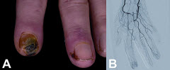
(BMJ) - A 46-yo woman w/ MS presented w/ discolored fingers, ulcerations, and necrosis in her left hand. Vasculitis antibodies and complement were normal. Angiography showed distal vasoconstriction. What drug was implicated in her Raynaud phenomenon?
Teriflunomide
Natalizumab
Glatiramer acetate
Interferon β1b
Fingolimod
You are correct. Although rare, Raynaud phenomenon and vasoconstriction are recognized side effects of interferon β, which the pt had started 2 ½ yrs prior. Treatment w/ IV prostaglandin E1 and prostacyclin analogues was attempted, but marked improvement occurred only after interferon was discontinued and glatiramer acetate started.
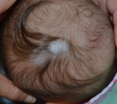
(BMJ) – An otherwise healthy 2-mo-old infant presented w/ 2 adjacent oval patches on the scalp, 2 x 3 cm in diameter, covered by a thin, atrophic membrane. The larger lesion exhibited the “hair collar sign.” MRI of the brain was normal. What is the diagnosis?
Cradle cap
Histiocytosis X
Atopic dermatitis
Tinea capitis
Aplasia cutis congenita (ACC)
You are correct. Aplasia cutis congenital (ACC) is a rare congenital malformation characterized by well-circumscribed absence of skin. Although the scalp is the most typical site, the entire skin can be involved. The “hair collar sign” consists of a ring of long, dark, thick hair surrounding the atrophic alopecic patches on the scalp. ACC may be associated w/ encephaloceles, meningoceles, and heterotopic brain tissue. Affected infants should undergo MRI to exclude a transcranial extension of soft tissue, such as spinal dysraphism or a neuroectodermal defect. Small areas of ACC usually heal well w/ alopecic scars; larger defects may require surgical intervention to reduce risk of infection and bleeding.