By vgreene, 8 June, 2015
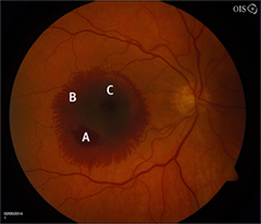
(BMJ)—A 67-yo woman presented w/ sudden onset of painless central vision loss in the right eye. Fundoscopy findings are shown. What is the dx?
Optic nerve drusen
Idiopathic perifoveal telangiectasia
Ruptured retinal artery macroaneurysm
Central retinal vein occlusion
Vitreous hemorrhage
You are correct. The fundoscopic examination of this pt showed a macular hemorrhage involving 3 tissue layers (trilaminar), characteristic of a ruptured retinal artery macroaneurysm: preretinal (obscuring the ruptured macroaneurysm—A); intraretinal (feathery border appearance—B); and subretinal (beneath the fovea, w/ retinal vessels coursing over—C). Because she presented promptly, the hemorrhage was successfully displaced after intravitreal injection of tPA and sulfur hexafluoride gas, plus a short postop period of face-down head posturing. Retinal artery macroaneurysms classically occur in elderly women and are strongly associated w/ HTN.